Mammography is a low-dose x-ray study of the breasts. The key role of mammography is in identifying a site of breast cancer early in its development when it is very small and often a year of two before it is large enough to be felt as a lump. These small cancers have a much better response to treatment and often require much less surgical or drug treatment. Mammography detects approximately 2-3 times as many ‘early’ breast cancers as physical examination, and is the best method for the screening of breast cancer.
Today, using digital mammography, very low doses of radiation are used for mammography. Digital mammography equipment is extremely expensive and for this reason few private hospitals or clinics install the machines.
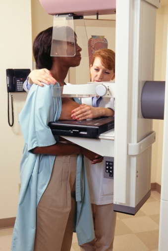
A typical mammogram consists of two views of each breast in which they are pressed firmly for a short time between two plates. The whole process takes only a few minutes and is not usually painful; some women do find it uncomfortable and if you do please tell the radiographer performing the examination.
Why do my breasts need to be compressed during mammography?
Breast compression is necessary to flatten the breast so that the maximum amount of tissue can be imaged and examined. Breast compression may cause some discomfort, but it only lasts for a brief time during the mammography procedure. Patients should feel firm pressure due to compression but no significant pain. If you feel pain, please inform the radiographer. The breast should only be compressed two to four times per breast for a few seconds each time.